By Preetesh Mistry, September 2023
Introduction
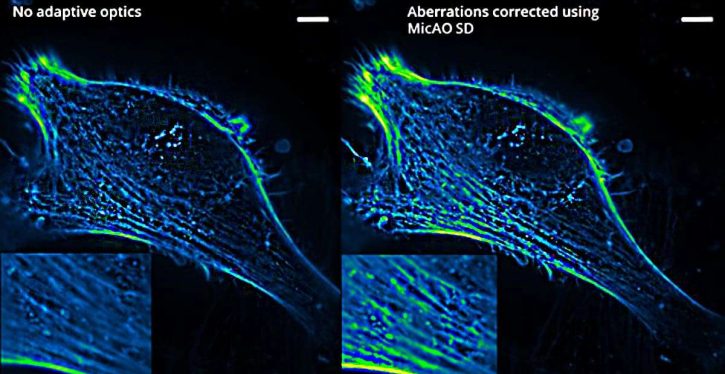
Optical components used within microscopes, such as objectives, lenses, and mirrors, as well as the biological samples being examined can induce aberrations. Imagine Optic has developed a number of solutions using adaptive optics to correct those aberrations and improve the contrast and resolution of acquired images for various microscopy modalities.
Advanced use of adaptive optics enables wavefront engineering and brings improvement to areas of imaging such as 3D single-molecule super-resolution at the cover slip as well as deeper in the biological sample. Aberrations can be efficiently corrected by using adaptive optics, such as the MicAO 3DSR from Imagine Optics. This is an adaptive optics device containing a Shack-Hartman-type wavefront sensor and continuous membrane deformable mirror. By correcting the spherical aberration, we can obtain perfectly symmetrical point spread function (PSF) along the Z-axis at a depth reaching 50µm in the sample. Following the aberration correction, the MicAO 3DSR can apply variable amounts of astigmatism for three-dimensional imaging.


Multiphoton Microscopy
In multiphoton microscopy, adaptive optics are implemented on the excitation pathway and can significantly boost the fluorescence signal from a biological sample. Correction of the system as well as the sample-induced aberrations improves the focusing ability of the microscope, with a typical gain of 3 to 5 times the two-photon effect.
The implementation of the adaptive optics starts with a determination of the optical aberrations present and then deploying a deformable mirror to correct them. The increase in fluorescence signal can allow for much deeper layers of the sample to be reached. It can also allow for a decrease in the intensity of the excitation laser which helps to prevent sample bleaching and minimises photo-toxicity effects.
SPIM / Light Sheet Microscopy
Selective Plane Illumination Microscopy (SPIM) or light sheet microscopy is currently the fastest imaging technique suitable for live imaging of voluminous biological samples. The samples and capillary, typically used as a sample holder, can induce aberrations that limit the optical quality of the imaging system. Adaptive optics have been proven to be highly effective at correcting these kinds of aberrations. The resolution and contrast of the image can be drastically improved which enables you to reach deeper layers into the biological sample.
Light sheet excitation is also becoming an increasingly popular technique in single molecule localisation microscopy (SMLM), where it excites fluorescence in only a thin section of the sample instead of in the whole volume. For example, with a specially designed sample holder, the single objective SPIM technique (referred to as soSPIM) uses the same objective lens to send the light-sheet excitation to the sample and to collect the emitted fluorescence signal. This results in an increased signal-to-noise ratio, allowing the user to reach deeper layers of the sample, where maintaining image quality would not be possible without adaptive optics aberration correction.

Figure 1. (A) Schematic representation of the soSPIM imaging system combined with the MicAO adaptive optics system from Imagine Optic for system and sample induced aberration correction, enabling in-depth 3D single molecule imaging. (B) Representation of the acquired volume 12 µm above the coverslip surface with an objective depth of focus of 1µm. (C) Raw single molecule DNA-PAINT data illustrating the 60 nm RMS astigmatism induced with the deformable mirror of the MicAO system in order to allow for 3D single molecule localization. (D) Depth-coded projection of the localization acquired within the volume shown in (B) of the protein Lamin-B1, demonstrating the 3-D shape of the nuclear envelope in non-adherent cells. * soSPIM was jointly developed by CNRS, University of Bordeaux and the National University of Singapore by Rémi Galland, Gianluca Grenci, Vincent Studer, Virgile Viasnoff and Jean-Baptiste Sibarita. https://doi.org/10.1038/nmeth.3402 This collaborative work between Imagine Optic and the Quantitative Imaging of the Cell group at the Interdisciplinary Institute for Neuroscience was possible thanks to the following funding: Cifre, ANR, soSPIM.
Confocal and Spinning Disk Microscopy for Live Cell Imaging
Scanning spinning disk confocal microscopy are popular imaging techniques in cellular biology for fixed cell and in-vivo imaging. In these modalities, the use of adaptive optics enables the user to increase the fluorescence signal and image contrast, especially when a high-magnification oil immersion objective is used to image water-based biological samples.
Fixed biological samples, primarily imaged using scanning confocal microscopy, induce complex aberrations which distort the image and reduce resolution and sharpness. The use of an adaptive optical element increases the signal-to-noise ratio to combat this distortion and reduction in sharpness. When imaging deeper into biological samples with the spinning disk technique, correction of aberrations using adaptive optics more than doubles the fluorescence signal. This also allows the PSF to be uniform in various parts of the sample, which dramatically improves the quality of image deconvolution when post-processing the data.
Further Information
MICAO 3DSR Adaptive Optics system Datasheet
Application note: Deep 3D PALM/STORM imaging combining depth and highest resolution
Application note: 3D dual-colour PALM/dSTORM imaging of centrosomal proteins with nanometric resolution
Application note: Quasi-isotropic nanometric 3D resolution in PALM/STORM
Application note: Temporal stabilisation module inside MICAO 3DSR